

美國病理學會是國際公認最具公信力的臨床醫學檢驗室認證機構。該機構定期進行實驗室調查和比較,以促進實驗室檢驗的品質提升。
在 2010 年發表於 Archives of Pathology & Laboratory Medicine 期刊的文獻 1 中,進行了對 2006 年 656 家美國病理學會細胞學實驗室的子宮頸抹片方法學的分析。
研究結果顯示:
1. 越來越多的細胞學實驗室將抹片技術從傳統抹片轉換為液態抹片 (LBP)
- 雖然仍有超過 60% (397/656) 的細胞實驗室同時使用兩種方法學,但只選用傳統抹片的實驗室已經從 24% 下降到 7%;相反,選用液態抹片作為唯一子宮頸癌篩檢方法的實驗室則增加了近 3 倍,由 9.3% 提升至 25.5%。
2. ThinPrep 為子宮頸液態抹片的首選
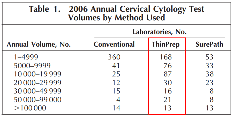
- 如表 1,子宮頸抹片年總量 5000 件以上的中大型細胞學實驗室選用 ThinPrep Pap Test 的比例最高。
- 根據 656 間實驗室的回覆數據統計,各種抹片方法學的平均使用率分別為:傳統抹片 7%;ThinPrep 57.2%;SurePath 19.6%。
3. ThinPrep 的鱗狀上皮異常檢出率比傳統抹片更高
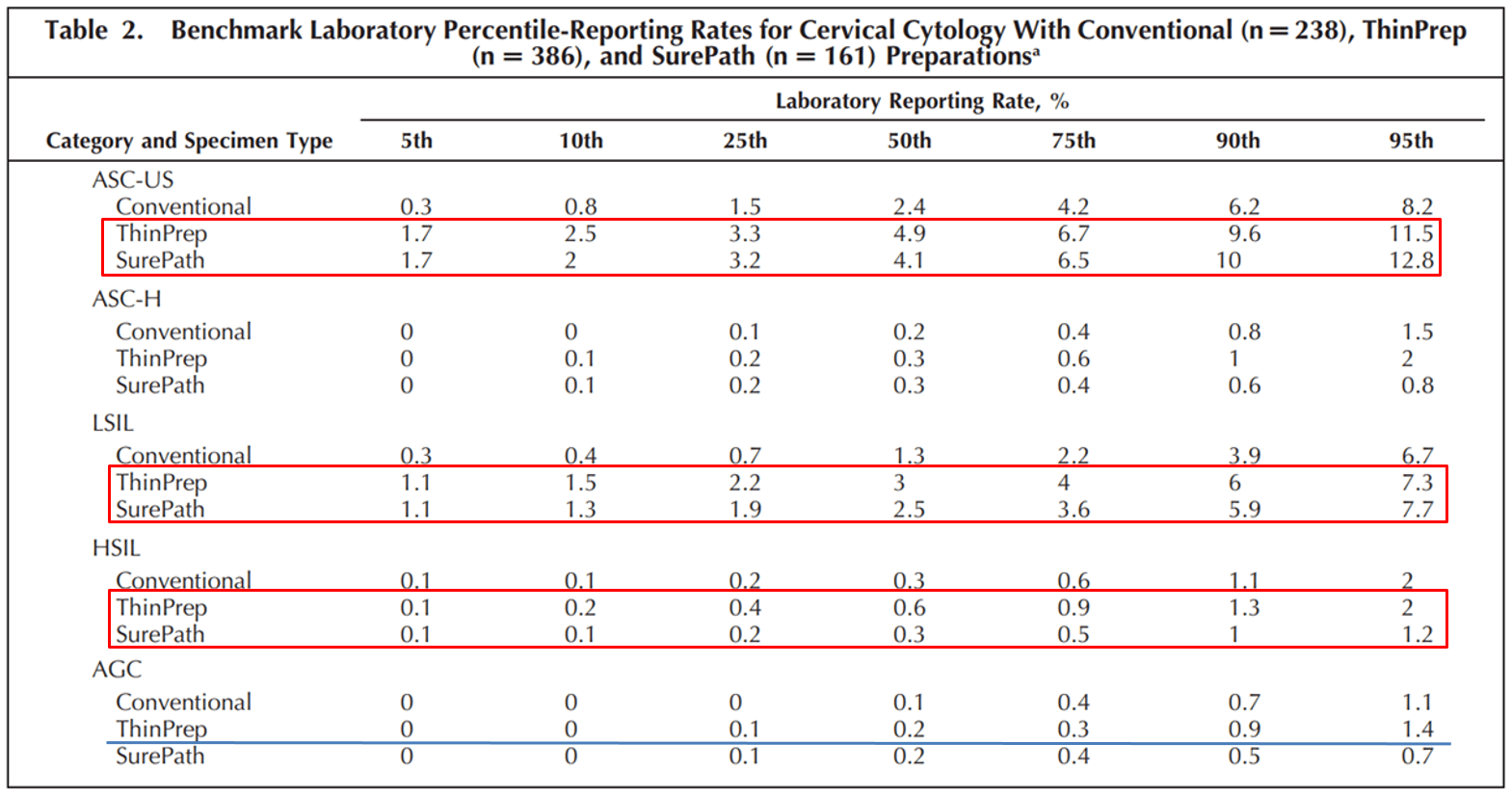
- 如表 2,液態抹片在檢出 ASC-US、LSIL 和 HSIL 的表現優於傳統抹片,並且具有統計學上的顯著性 (p <0.05)。
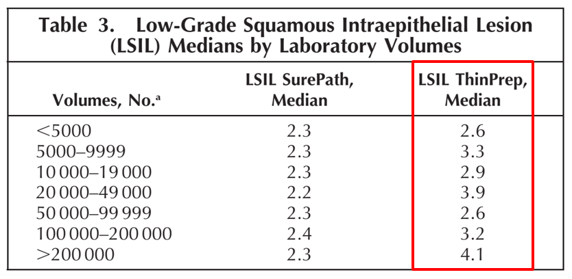
- 在鱗狀上皮異常的診斷類別,ThinPrep 幾乎在所有百分比群組中具有最高的檢出率。如表 3,ThinPrep Pap Test 在 656 間實驗室的 LSIL 檢出率表現優於其他方法。
- 在診斷腺體上皮異常 (AGC) 的類別中,ThinPrep 的檢出率也最高。ThinPrep Pap Test 是唯一獲得美國 FDA 核准,具有促進子宮頸腺癌檢出率的抹片方法2-7。
根據上述研究統計資料,ThinPrep 液態抹片相較於傳統抹片,在鱗狀上皮異常和腺體病灶檢出方面具有更高的檢出率,因此已成為液態抹片的主要品牌。此外,ThinPrep Pap Test 檢體還可同時進行人類乳突病毒(HPV)檢測,co-testing 兼具高靈敏度和高特異性,提供完整的子宮頸癌篩檢效能,對女性健康具有重要的防護作用。
Co-testing 資訊請見:子宮頸癌怎麼篩?CAP 專家:只用 HPV test 篩檢子宮頸癌風險高 建議採 co-testing 方案
欲了解詳細資訊,歡迎聯繫產品專員 陸詩羽,連絡電話: 02-2246-7799 #805
參考資料:
- Practices of Participants in the C ollege of American Pathologists nterlaboratory Comparison Program in Cervicovaginal Cytology, 2006. Arch Pathol Lab Med—Vol 134, March 2010
- Histologic follow-up of atypical endocerv cal cells. liquidbased, thin-layer preparation vs. conventional pap smear. Acta Cytol. 2002;46:453-457.
- ThinPrep Pap Test promotes detection of glandular lesions of the endocervix. diagn Cytopathol. 2000;23:19-22.
- ThinPrep Pap Test: Performance biopsy follow-up in a university hospital. Cancer.1999;87:105-112.
- detection of endometrial adenocarcinoma with the ThinPrep Pap Test. diagn Cytopathol. 2000;23:260-265.
- ThinPrep Pap Test: Accuracy for glandular disease. Acta C tol. 1999;43:81-85.
- ThinPrep detection of cervical and a typical endocervical cells: a retrospective cohort study. Cancer Cytopathol. 2002;96:338-343